Causes and Diagnoses
Causes and Diagnoses of Crohn's Disease
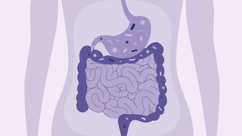
While the exact cause of Crohn's disease is unknown, the disease process is well understood. It is an autoimmune disease, meaning the immune system attacks healthy tissue in response to bacteria that are normally present to aid in digestion. The immune response creates chronic inflammation that leads to ulcers and thickening of the lining of the digestive system.
Recent research has shown that several factors are involved:
- Environmental factors: Crohn's disease is found more frequently in developed countries, urban areas and cooler climates.
- Genetics: A family member of someone with Crohn's disease is significantly more likely to develop the condition.
- Ethnicity: Crohn's disease is most common among people of east European descent, including Ashkenazi Jews. The rate of Crohn's disease is increasing in African-American populations.
- Stress: While not a cause, stress may aggravate Crohn's symptoms.
- Diet: Certain foods may trigger symptoms in some individuals.
Diagnosing Crohn's disease
Several tests can aid your physician in diagnosing Crohn's disease. These tests can also be repeated from time to time to evaluate the progress of the disease. They include:
- Blood test: Lab tests can identify infection, antibodies, anemia, and vitamin and mineral deficiencies.
- Fecal blood test: This test shows if there is blood in your stool, indicating inflammation.
- X-ray: Traditional X-ray images can indicate the severity of the inflammation and scarring, and show if there are any blockages.
- White blood cell scan: White blood cells, which combat infection, are isolated from your blood sample, tagged with radioactive material, and injected into your body. Later, a nuclear scanner is used to identify where the white blood cells have gathered.
- CT scan: A computed tomography (CT) scan combines X-ray and computer technology to produce detailed cross-sectional images of your intestines.
- Colonoscopy: An endoscope (long, flexible tube) with a lighted camera goes through the colon, allowing your physician to view the lining. A sigmoidoscopy uses the same technology but examines only the sigmoid colon (the lower third).
- Endoscopic ultrasound (EUS): While you are under mild sedation, an endoscope with an ultrasound probe is inserted in your colon to create more precise images than may be available using external ultrasound.
- Endoscopic retrograde cholangiopancreatography (ERCP): An endoscope goes through your esophagus, stomach and into the beginning of your small intestine. X-ray technology and contrast dye create clear images of the bile ducts.
- Magnetic resonance imaging (MRI): This test uses a magnetic field and radio waves to create pictures of your digestive tract.