


How To Read an Electrocardiogram (ECG)
A Cardiologist Explains What the Waves Can (and Can’t) Tell You
Updated October 2026
An electrocardiogram (ECG) is a quick, painless test that records your heart’s electrical activity.
It’s one of the most common tools physicians use to check how your heart is working, and it often takes just a few minutes. You may also hear this test called an EKG. ECG and EKG refer to the same thing.
Every cardiac complaint begins with an ECG.— Micah J. Eimer, MD
“Every cardiac complaint begins with an ECG, whether you have chest pain, shortness of breath or an irregular heartbeat,” says Micah J. Eimer, MD, a cardiologist at Northwestern Medicine and a clinical assistant professor of Medicine at Northwestern University Feinberg School of Medicine.
It takes years of training to fully interpret an ECG, but understanding the basics can help you better understand your heart health and participate in decisions about your care.
What Does Your Heart Look Like?
To understand what an ECG shows, it helps to know what your heart is doing.
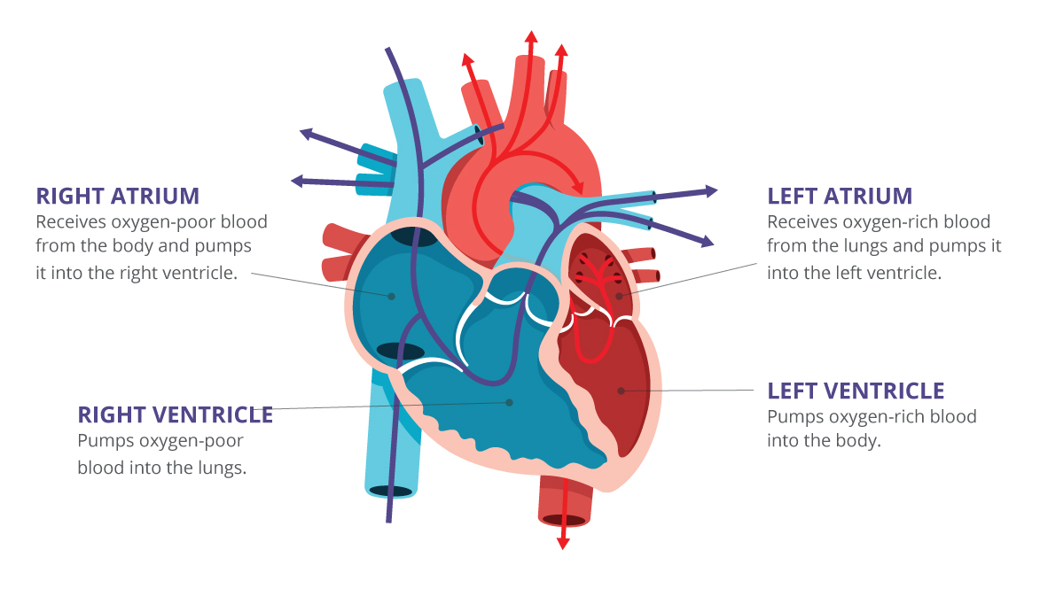
Your heart has four chambers: a right and left atrium on top, and a right and left ventricle below.
The right side collects and sends oxygen-poor blood to your lungs, where it picks up oxygen. The left side receives the oxygen-rich blood and pumps it out to the rest of your body.
Your heart repeats this cycle about 100,000 times a day, moving roughly 2,000 gallons of blood.
For the chambers to work in the right order, your heart relies on electrical signals to tell each one when to squeeze. An ECG records those signals.
What the Waves Mean
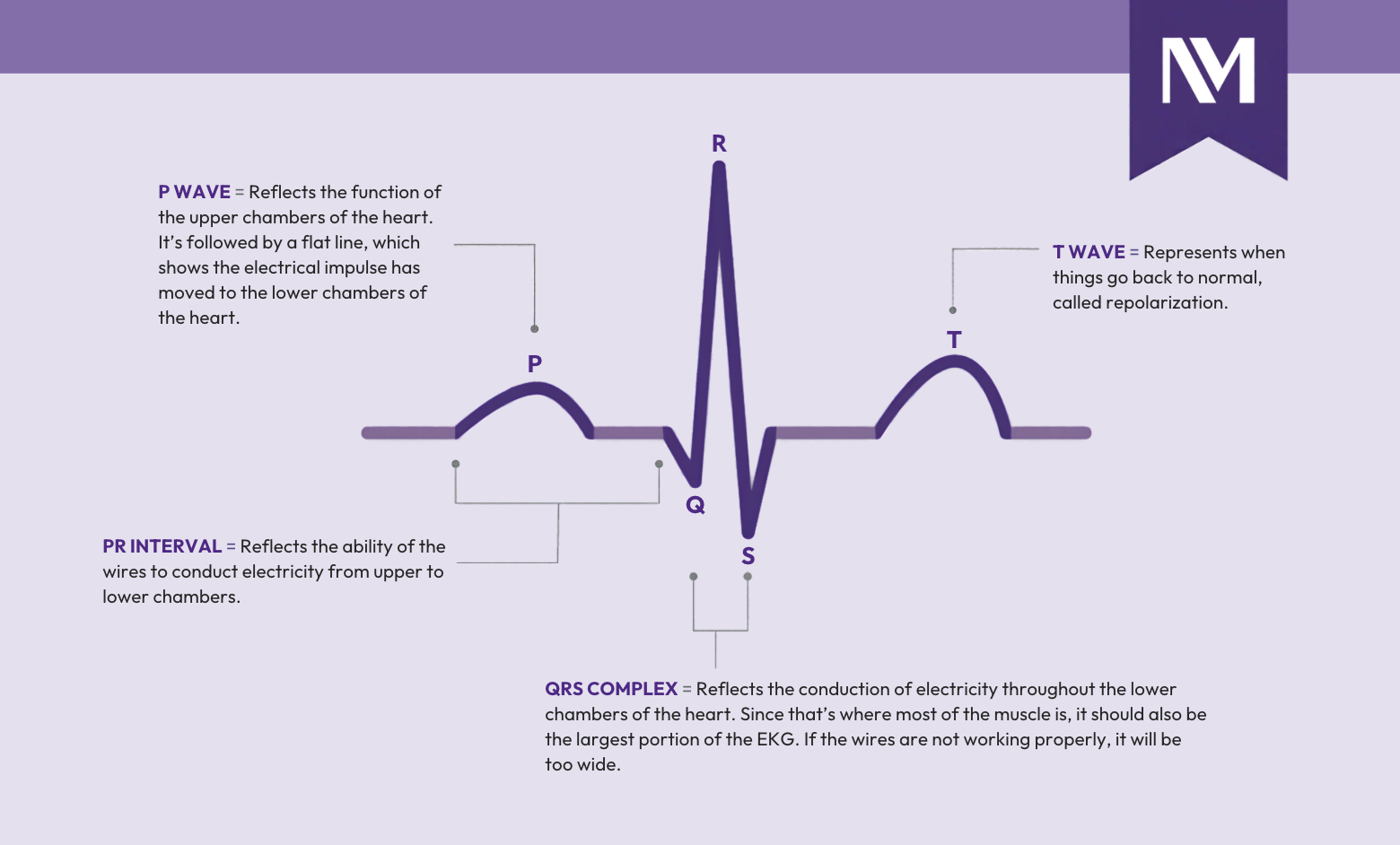
On an ECG, each heartbeat appears as a repeating pattern of waves. Each part of that pattern reflects a different step in the electrical signal that moves through your heart.
- P wave is the first small bump. It reflects electrical activity in the upper chambers of your heart.
- PR interval is the stretch between the P wave and the tall spike. It shows the signal traveling from the upper chambers to the lower chambers.
- QRS complex is the tall, sharp spike. It shows electricity moving through the lower chambers, which are the part of the beat that pumps blood out to your lungs and body.
- T wave is the rounded wave that follows. It reflects your heart’s electrical system resetting before the next beat.
Your physician looks at the size, shape and spacing of these waves. Changes in any of them can point to a rhythm problem, strain on the heart or an area of injury.
What an ECG Can Detect
An ECG can help detect:
- An irregular heartbeat (arrhythmia)
- A heart rate that is too fast or too slow
- Signs that the heart may be enlarged or overworked
- Signs of a current or previous heart attack
- Changes that may suggest an injury to the heart muscle
Some consumer smartwatches and wearable devices can flag an irregular rhythm or resting heart rate, but they don’t replace a medical-grade ECG. If a device alerts you, talk with your doctor.
What’s a Normal Heart Rate?
Heart rate is one of the easiest ECG results to understand. For most adults, a normal resting rate falls between 60 and 100 beats per minute, though well-trained athletes often run lower and are perfectly healthy.
What matters most is whether your rate is consistently too fast or too slow, especially alongside dizziness, fatigue or palpitations.
What an ECG Can’t Tell You
An ECG gives your care team a lot of information, but it doesn’t show everything. It does not directly show blocked coronary arteries or how well blood is flowing through your heart.
A normal ECG also doesn’t always rule out a heart problem. Some conditions affect the heart only occasionally and may not appear during the test.
Depending on your symptoms, your physician may recommend additional tests, such as:
- An echocardiogram, which uses sound waves to create images of your heart’s structure and pumping function
- A stress test, which records your heart’s activity while you exercise
- A heart rhythm monitor, such as a Holter monitor, which you wear for a day or longer to capture irregular beats that come and go
- Cardiac imaging, such as a computed tomography (CT) or magnetic resonance imaging (MRI) scan, which shows the heart and blood vessels in greater detail
When Might You Need an Electrocardiogram?
Your physician may recommend an ECG if you experience:
- Chest pain
- Heart palpitations
- Shortness of breath
- Dizziness
- Fainting
- An irregular heartbeat
An ECG may also be performed before surgery or other medical procedures to help evaluate your heart health.
What To Expect During an ECG
During an ECG, a member of your care team will place small adhesive electrodes on your chest and sometimes your arms and legs.
The test is:
- Painless
- Noninvasive
- Quick, often taking only a few minutes
The electrodes record your heart’s electrical signals. You won’t feel any electrical current during the test.
When To Seek Immediate Medical Attention
Some heart problems are life-threatening and need treatment right away. Call 911 or seek emergency care right away if you have:
- Chest pain or pressure
- Shortness of breath
- Symptoms of a heart attack or stroke
Do not assume symptoms are harmless because you previously had a normal ECG.
How AI Is Helping Spot Heart Problems Earlier
Heart disease often goes undiagnosed until it causes serious complications. To catch it sooner, Northwestern Medicine Bluhm Cardiovascular Institute uses artificial intelligence (AI) to take a closer look at ECG results.
The technology analyzes ECG recordings for patterns linked to atrial fibrillation (AFib) and atrial flutter, which are irregular rhythms that can raise your risk of stroke. It’s designed to flag patients who may be likely to develop AFib within the next year, before symptoms appear.
“The algorithm reviews outpatient ECGs and results go to the physician who ordered the test,” says Dr. Eimer. “The algorithm doesn’t make the diagnosis. It flags patients who deserve a closer look, and the physician decides what comes next.”
Learn more about taking care of your heart.
Featured Experts



 Chrome
 Edge
Edge Safari
 Firefox