Treatment
Treatment for Melanoma
Melanoma care with Northwestern Medicine Dermatology
Staging
Melanoma is one of the more dangerous types of skin cancer. It is responsible for the greatest number of skin cancer deaths. According to the American Cancer Society, there are over 106,000 new melanomas diagnosed each year, as well as over 7,000 deaths.1 While this can stem from existing moles, more often, it shows up suddenly on healthy skin. Once a melanoma develops, it may spread to other areas of the body. This serious skin cancer is highly curable if caught early. This is why early diagnosis and treatment are critical.
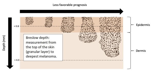
Your clinician uses staging to help decide your treatment. Breslow thickness is the depth of the primary tumor’s vertical invasion into the skin. This is the most important factor your physician uses to determine your prognosis.2
| T Category | Breslow Thickness (millimeters) | Ulceration Status |
| T1a | less than or equal to 0.8 | without |
| T1b | 0.8 - 1.0 | with or without |
| T2a | greater than 1.0 - 2.0 | without |
| T2b | greater than 1.0 - 2.0 | with |
| T3a | greater than 2.0 - 4.0 | without |
| T3b | greater than 2.0 - 4.0 | with |
| T4a | greater than 4.0 | without |
| T4b | greater than 4.0 | with |
*The presence of ulceration (a break in the skin causing both loss of the epidermis and exposure of the underlying dermis) automatically upgrades the sub-category from “a” to “b.”3
Melanoma is usually staged using the American Joint Committee on Cancer’s TNM staging system. Staging a tumor helps your physician understand the prognosis or outlook for what the disease course may look like. The TNM stage also helps your physician decide the best course of treatment and follow-up monitoring.
There are three main components of the TNM staging system:
- T – Primary Tumor. This is calculated based on how deep (in millimeters) the tumor cells reach into the layers of the skin. It is recorded as the Breslow thickness as well as the presence or absence of ulceration.
- N – Nodal involvement. This component is based on if any tumor cells are detected in lymph nodes next to the main site where the melanoma was found.
- M – Metastasis. This component measures any spread of the tumor to distant body sites.
Lower numbers in each category correspond to a more favorable prognosis. For instance, a melanoma stage T1aN0M0 has a better prognosis than a melanoma stage T2bN1M1. Together, the combination of the three numbers forms a clinical stage. Your physician uses this to guide how to treat the melanoma and monitor for recurrence or progression of disease.
We also use other staging tools such as the online AJCC calculator and molecular staging tools involving gene expression profiling.
Treatment
The melanoma stage has a significant role in determining treatment. After a melanoma is confirmed via biopsy, your dermatologist will make treatment suggestions based on the melanoma’s stage, your genetic profile and your overall health.