PET CT
Positron Emission Tomography/Computed Tomography (PET/CT)
 Positron emission tomography—computed tomography (PET/CT) provides highly detailed images that can help fine-tune a diagnosis for cancer, heart disease or brain disorders.
At Northwestern Medicine, you’ll find the most current advances in PET/CT along with board-certified radiologists* and licensed technologists to guide your exam. PET/CT exams are performed in a convenient, comfortable outpatient setting.
How PET/CT works
PET/CT is a diagnostic imaging tool that combines two scan techniques: positron emission tomography (PET) and computed tomography (CT). It uses a small dose of radiation to create detailed images of structures and functions inside your body and can detect problems that don’t show up on other types of diagnostic imaging exams.
- Computed tomography (CT or CAT scan): CT is a noninvasive diagnostic imaging procedure that uses a combination of X-rays and computer technology to produce both horizontal, or axial, images (often called slices) of the body. A CT scan shows detailed images of any part of the body, including the bones, muscles, fat and organs.
- Positron emission tomography (PET): PET is a specialized radiology procedure used to examine various body tissues to identify certain conditions. PET may also be used to follow the progress of the treatment of certain conditions. PET is a type of nuclear medicine procedure. This means that a tiny amount of a radioactive substance, called a radionuclide (a radiopharmaceutical or radioactive tracer), is injected into the body during the procedure to assist in the examination of the tissue under study. A special type of camera can then detect the radioactivity in the body.
How PET/CT is used
Physicians use PET/CT in many ways:
- To detect cancers and assess the effect of cancer therapy
- To diagnose heart disease and gauge heart muscle damage after a heart attack
- To examine the brain for people with specific brain disorders
About your exam
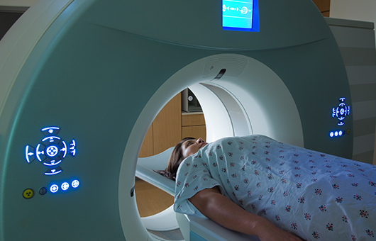
During a PET/CT scan, you are first injected with a radioactive substance. Lying on a flat table, you move slowly through a doughnut-shaped machine that detects positrons, tiny particles given off by the radioactive material. The machine takes a series of thin "slice" images, which are then assembled to create a three-dimensional image of your body.
A radiologist will interpret results of your exam and report them to your physician. PET/CT scanning is provided on an outpatient basis.